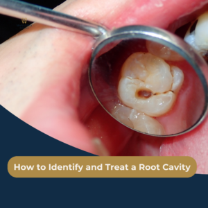
When most people imagine a tooth cavity, they picture a visible dark spot on the chewing surface of a molar or a discolored gap between their front teeth. However, one of the most destructive forms of tooth decay occurs out of sight: root cavities (also known as root caries). These lesions form at or below the gumline on the root surface of the tooth, operating silently until significant structural damage has already taken place.
Why Root Surfaces Are Uniquely Vulnerable
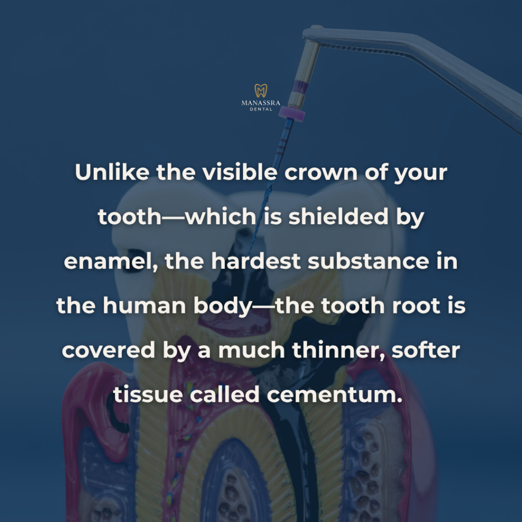
Unlike the visible crown of your tooth—which is shielded by enamel, the hardest substance in the human body—the tooth root is covered by a much thinner, softer tissue called cementum. Beneath the cementum lies dentin, a porous structure containing microscopic tubules that lead directly to the tooth’s nerve center (the pulp). Once gum tissue recedes due to age, aggressive brushing, or periodontal disease, this delicate root surface becomes exposed to oral bacteria. Because dentin demineralizes at a much lower acid threshold than enamel, decay on exposed roots spreads more than twice as fast as standard crown decay.
The Strategic Solution at Manassra Dental
Because sub-gingival decay progresses rapidly toward the dental nerve, early clinical identification is critical. At Manassra Dental in San Jose, our diagnostic approach uses advanced digital imaging and specialized clinical screening to detect subgingival lesions before they require invasive endodontic therapy or tooth extraction. Understanding how root cavities develop, recognizing subtle early warning signs, and knowing your treatment options will help you safeguard your smile for years to come.
Why Root Cavities Form
Root cavities do not occur on healthy, fully covered teeth. They require a specific combination of biological shifts and physical conditions to take root.
The Primary Drivers of Sub-Gingival Decay
- Gingival Recession (Receding Gums): The foundational prerequisite for a root cavity is an exposed root surface. Gum tissue can pull back due to chronic periodontitis, natural aging, hormonal shifts, orthognathic stress, or the use of a hard-bristled toothbrush.
- Xerostomia (Chronic Dry Mouth): Saliva is your mouth’s natural defense mechanism; it washes away food debris, neutralizes bacterial acids, and supplies essential minerals (calcium and phosphate) to remineralize enamel and dentin. Many prescription medications—including blood pressure drugs, antihistamines, and antidepressants—reduce salivary flow, dramatically increasing the risk of root caries.
- Plaque Accumulation at the Margin: When plaque settles in the narrow groove where the tooth meets the gumline (the gingival sulcus), oral bacteria consume dietary sugars and excrete harmful acids that rapidly dissolve exposed cementum and dentin.
- Complex Restorative Margins: Existing crowns or fillings placed near the gumline can develop micro-gaps over time, creating ideal harbors for recurrent sub-gingival bacteria.
Catching Decay Early
Recognizing a root cavity in its earliest stages requires paying close attention to minor sensory shifts in your mouth. Because root decay occurs near the nerve, initial symptoms are often sensory rather than visual.
Key Warning Signs to Watch For
- Localized Temperature Sensitivity: A sharp, lingering twinge when consuming hot coffee, ice water, or sweet foods, specifically near the gumline.
- Tactile Tenderness During Brushing or Flossing: Feeling a “sticky” or tender sensation when your toothbrush bristles catch near the base of a tooth.
- Visible Discoloration Along the Gumline: Soft, yellowish-brown or dark brown spots appearing where the tooth meets the gum.
- Notching or Grooving: Feeling a physical indentation with your tongue or fingernail along the base of the tooth.
How Manassra Dental Diagnoses Sub-Gingival Decay
Diagnosing root caries requires specialized tools, as standard visual checks often miss decay hiding beneath the soft tissue margin:
- Low-Radiation Digital Radiographs (Bitewing X-Rays): X-rays penetrate beneath the gumline to reveal hidden interproximal root lesions and assess how close the decay has traveled toward the pulp.
- Tactile Exploration with Tactile Probes: Our clinical team uses delicate dental instruments to assess the texture of exposed dentin; active decay feels soft and leathery compared to sound, hard tooth structure.
- Intraoral Digital Cameras: High-magnification digital photography allows us to project high-definition images onto overhead monitors so you can see exact areas of recession and surface breakdown alongside your doctor.
Clinical Treatment Options at Manassra Dental
The appropriate treatment for a root cavity depends on how deeply the decay has penetrated the root tissue and whether the dental pulp remains healthy.
Treatment Spectrum for Root Caries
- Remineralization & Topical Fluoride Therapy (Early-Stage Non-Invasive): If a root lesion is detected in its earliest stage before a physical cavity (cavitation) forms, high-strength prescription fluoride varnishes or silver diamine fluoride (SDF) can halt bacterial growth and re-harden the dentin surface without drilling.
- Composite Resin & Glass Ionomer Restorations (Moderate Decay): For established cavities that have formed a physical notch, the decayed tissue is gently removed. We restore the root contour using Glass Ionomer Cement (GIC) or tooth-colored composite resin. Glass ionomer is particularly effective below the gumline because it bonds exceptionally well to dentin and continually releases fluoride into surrounding tissues to prevent future decay.
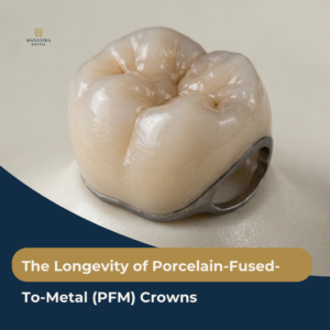
- Root Canal Therapy & Crown Placement (Advanced Internal Decay): If sub-gingival decay reaches the pulp chamber, the internal nerve becomes infected. In these cases, Dr. Manassra performs a precise root canal procedure to clear the infection, seal the internal canals, and place a custom crown to reinforce the tooth’s structural integrity.
- Periodontal Crown Lengthening: If decay extends deep beneath the gumline, a minor surgical contouring of the tissue (crown lengthening) may be performed to expose clean, healthy tooth structure for a stable, long-lasting restoration.
Frequently Asked Questions About Root Cavities
Yes. Because the root forms the narrow structural anchor of the tooth, unmanaged decay that wraps around the root circumference (known as circumferential caries) hollows out the solid dentin base. Without timely restoration, standard chewing pressure can cause the natural crown of the tooth to snap off completely at the gumline.
While receded gum tissue usually does not grow back naturally, you can protect exposed roots by switching to a soft-bristled electric toothbrush, using a prescription high-fluoride toothpaste (such as 5000 ppm sodium fluoride), staying hydrated, and using alcohol-free antibacterial mouth rinses. Regular hygiene cleanings and checkups at Manassra Dental ensure any early softening is treated immediately.
Schedule Your San Jose Evaluation
Root cavities are a silent but manageable threat to your long-term health. By combining daily oral hygiene with professional preventative care, you can protect exposed root surfaces, stop decay in its tracks, and avoid complex dental procedures.
At Manassra Dental, we are dedicated to providing compassionate, precise restorative care tailored to the San Jose community. Whether you are experiencing new tooth sensitivity near the gumline or are due for a routine clinical examination, our team is here to help you preserve your natural teeth for a lifetime.